
Articles
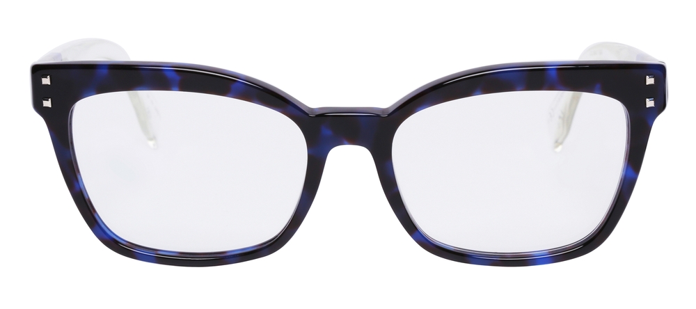
Technology continues to blur the lines between style and functionality - and Nuance Audio Glasses are a perfect example of this evolution. Designed to look like everyday eyewear, these innovative glasses feature built-in hearing enhancement technology that helps users hear conversations clearly while maintaining natural, effortless vision.
What Are Nuance Audio Glasses?
Nuance Audio Glasses are smart eyeglasses designed to provide both vision correction and enhanced hearing. Developed by Nuance Hearing, these glasses discreetly integrate directional microphones, miniature speakers, and Bluetooth connectivity into the frame - allowing wearers to improve their hearing experience without traditional hearing aids. By combining hearing technology with fashionable eyewear, Nuance Audio Glasses offer a seamless, everyday solution for individuals who want better hearing support without drawing attention to their device.
How Nuance Audio Glasses Work
The technology behind Nuance Audio Glasses focuses on selective sound amplification and speech clarity. The glasses use beamforming microphones to focus on voices in front of the wearer while reducing background noise. The enhanced sound is then transmitted through tiny speakers near the ears, allowing users to hear clearly without the occlusion effect common with in-ear devices.
This open-ear design keeps the ear canals unobstructed, promoting natural hearing while boosting important speech frequencies - ideal for conversations in noisy environments like restaurants or social gatherings.
For many individuals, dry eye disease is an ongoing struggle marked by irritation, burning, watery eyes, and inconsistent vision. While over-the-counter drops may offer short-term relief, they often fail to provide lasting hydration. In many cases, the issue isn’t a lack of tear production - it’s that tears drain away too quickly.
One innovative option designed to provide longer-term relief is LACRIFILL®, a modern approach to punctal occlusion that helps preserve your natural tears and restore balance to the ocular surface.
What Is LACRIFILL® Canalicular Gel?
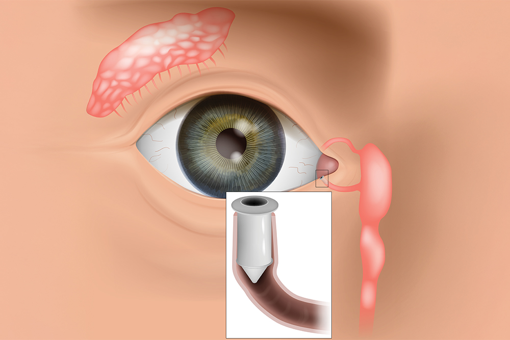
LACRIFILL® is a soft, clear gel that is placed inside the eye’s natural tear drainage channels, known as the canaliculi. These tiny channels are responsible for carrying tears away from the surface of the eye and into the nasal cavity.
For individuals with dry eye disease, tears often drain away too quickly. When this happens, the eye does not stay properly lubricated, leading to dryness and irritation. LACRIFILL® works by gently filling part of the drainage pathway, slowing tear outflow so that your natural tears remain on the eye longer.
Unlike traditional artificial tears that supplement moisture from the outside, LACRIFILL® helps you retain the tears your body already produces. This treatment is FDA-cleared and has been clinically studied for safety and effectiveness in managing dry eye symptoms.
How Does LACRIFILL® Work?
Every time you blink, a thin layer of tears spreads across the surface of your eye. This tear film keeps your eyes smooth, hydrated, and protected from debris and infection. After serving their purpose, tears drain through small openings in the inner corners of the eyelids (called puncta), passing into the canaliculi and eventually into the nose. In dry eye patients, this drainage process may occur too rapidly, leaving the surface of the eye exposed and under-lubricated.
LACRIFILL® forms a soft gel barrier within the drainage system, reducing tear loss and improving moisture retention. By keeping tears on the eye longer, it supports a healthier tear film and enhances overall comfort.
The gel gradually biodegrades over time and is naturally reabsorbed by the body. Relief typically lasts several months, and the treatment can be repeated when necessary.
Benefits of LACRIFILL®
LACRIFILL® offers several advantages for patients struggling with persistent dry eye symptoms:
FDA-cleared and clinically evaluated for safety and performance
Quick, non-surgical procedure performed in-office
No downtime or recovery period required
Preserves your body’s natural tears rather than relying solely on artificial drops
Provides extended relief that can last for months
Biocompatible material designed to work harmoniously with the body
Customizable treatment approach based on your individual tear drainage system
For patients who are tired of frequent eye drop use, this longer-lasting solution can be life-changing.

In the realm of eye care, surgical co-management has emerged as a collaborative approach that aims to provide patients with comprehensive and seamless treatment. This concept involves the joint efforts of optometrists and ophthalmologists, each bringing their unique expertise to the table. By working together, these eye care professionals strive to enhance patient outcomes and deliver exceptional care.
The Role of Optometrist and Ophthalmologist in Surgical Co-Management
Surgical co-management is built upon the unique skill sets and areas of expertise of optometrists and ophthalmologists. By understanding their respective roles, you can appreciate the synergy that this collaborative approach fosters.
Optometrists are primary eye care professionals who specialize in the examination, diagnosis, and non-surgical treatment of vision disorders. Their responsibilities in surgical co-management include:
Performing comprehensive eye examinations and evaluations
Monitoring and managing pre-existing eye conditions
Providing pre-operative and post-operative care
Educating patients on surgical procedures and aftercare
Collaborating with ophthalmologists to ensure continuity of care
Ophthalmologists are medical doctors who specialize in the diagnosis, treatment, and surgical management of eye diseases and disorders. Their role in surgical co-management encompasses:
Evaluating patients' candidacy for surgical interventions
Performing complex surgical procedures
Providing specialized medical and surgical care
Collaborating with optometrists to ensure seamless patient care
Monitoring and managing post-operative complications
By combining the expertise of optometrists and ophthalmologists, surgical co-management ensures that patients receive comprehensive and coordinated care throughout their treatment journey.
How Surgical Co-Management Works
Surgical co-management is a well-orchestrated process that involves several key steps. Understanding how it works can help you navigate this collaborative approach with confidence.
Initial Evaluation: The process typically begins with an optometrist conducting a comprehensive eye examination. During this evaluation, the optometrist assesses the patient's visual needs, identifies any potential issues, and determines if a surgical intervention is necessary.
Referral and Consultation: If surgery is recommended, the optometrist refers the patient to an ophthalmologist for further evaluation and consultation. This step ensures that the patient receives specialized medical advice and a thorough assessment of their suitability for the proposed surgical procedure.
Pre-operative Care: The optometrist plays a crucial role in providing pre-operative care, which may include managing any existing eye conditions, ensuring the patient understands the surgical process, and addressing any concerns or questions they may have.
Surgical Procedure: The ophthalmologist performs the necessary surgical intervention, leveraging their specialized training and expertise in surgical techniques.
Post-operative Care: After the surgery, the patient's care transitions back to the optometrist, who closely monitors the recovery process and provides post-operative care and management. This may involve follow-up appointments, monitoring for any complications, and ensuring the patient adheres to the prescribed treatment plan.
Ongoing Collaboration: Throughout the entire process, the optometrist and ophthalmologist maintain open communication and collaborate closely. This ensures that the patient's care is seamless, and any concerns or issues are promptly addressed by the appropriate healthcare professional.

In September 2025, the FDA approved Stellest lenses for children ages 6 to 12 to correct and manage myopia, a major milestone for myopia care. These lenses have been successfully used in countries across Europe, Canada, and Asia since 2020, showing promising results in slowing eye growth associated with myopia.
The Importance of Early Myopia Management
Myopia typically begins in early childhood, often between the ages of 6 and 10, and tends to worsen quickly during the school years as children spend more time reading and using digital devices. The sooner it begins, the higher the risk that it will progress to more severe levels. Every increase in myopia level significantly raises the risk of eye health complications later in life, such as retinal detachment or glaucoma.
By beginning myopia management as soon as possible, parents can help slow eye growth and protect long-term vision. With innovative options like Stellest lenses, families now have effective, science-backed solutions to take control of their child’s myopia.
What Are Stellest Lenses?
Stellest lenses are a groundbreaking innovation in myopia control, developed by Essilor. They look like regular eyeglass lenses but are designed with specialized optical technology to both correct vision and slow the progression of myopia.

Thanks to the advancement of lens technology, glasses lenses are no longer a single, one size fits all solution. There are a variety of different lens types that can be used in glasses, giving patients greater flexibility and control over their vision than ever before.
Single Vision Lenses
Also known as monovision lenses, these lenses are designed to correct the wearer’s vision at just one distance, and have a single prescription covering the entire surface of the lens. They are most often recommended for people who are either nearsighted (myopia) or farsighted (hyperopia) and who need glasses for a specific activity, such as driving or reading.
Progressive Lenses
Progressive lenses are multifocal lenses that can correct a patient’s vision at different working distances, ranging from far distance to reading distance. However, rather than designating different areas on the lenses for different distances with visible lines separating them, progressive lenses have a gradual change so that the wearer can smoothly transition from one lens power to another.
Bifocal and Trifocal Lenses
As you may have guessed from the name, bifocal and trifocal lenses have either two or three lens powers depending on which type you choose. Bifocal lenses support distance vision in the top half of the lens, and near vision in the lower half. Trifocal lenses support distance vision in the top third of the lens, intermediate vision in the middle segment and near vision in the bottom third. Whichever variety you choose, you will see visible lines separating each segment.
Bifocal and trifocal lenses are recommended for patients who are near or farsighted, and those who develop presbyopia, which is the natural hardening of the eye lens, that occurs as we get older. Presbyopia makes it harder for the lens of the eye to adapt to focus at different distances.
Multifocal Lenses
Multifocal lenses are the alternative name given to bifocal, trifocal and progressive lenses.
Neuro-Optometric Rehabilitation
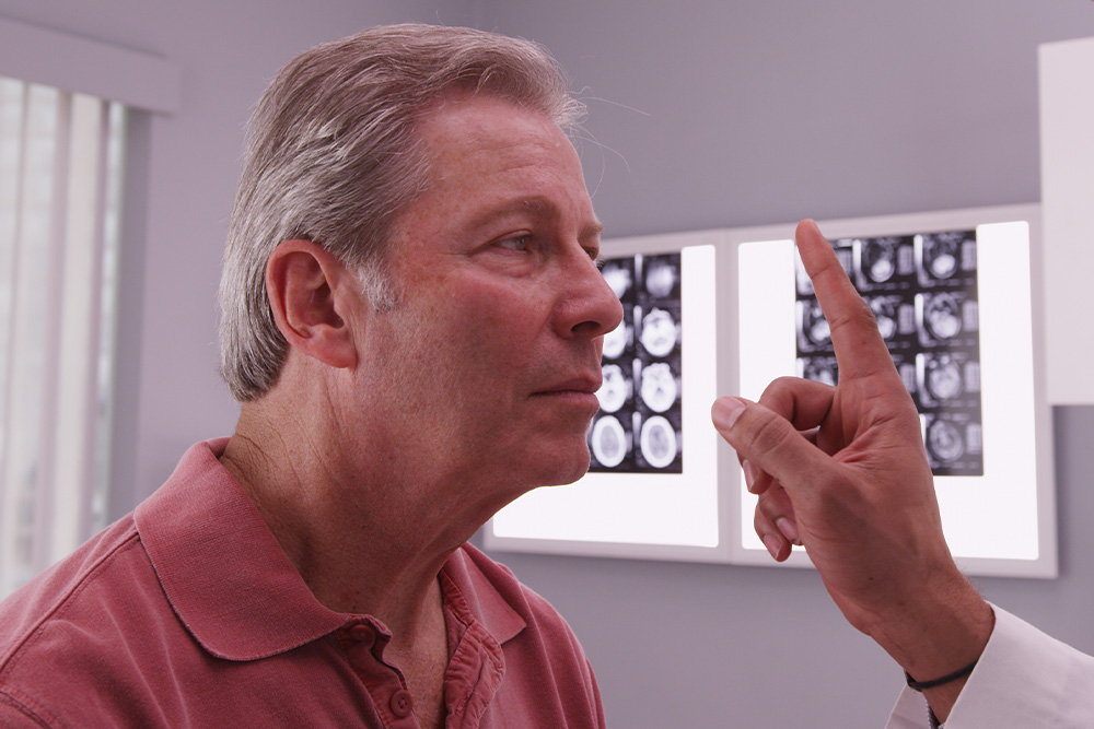
When most people think about vision problems, they picture issues with the eyes themselves. However, many visual difficulties actually begin in the brain. After a concussion, stroke, or other brain injury, the connection between the eyes and brain can be disrupted - causing symptoms that affect balance, reading, focus, and even daily comfort.
Brain Injuries and Vision
Your visual system is complex - about 70% of the brain is involved in vision processing. After a traumatic brain injury (TBI), concussion, or neurological event such as a stroke, the pathways that coordinate eye movement, focus, and perception can become impaired.
This can result in symptoms such as:
Blurred or double vision
Eye strain and headaches
Difficulty reading or focusing
Dizziness or balance problems
Light sensitivity
Problems with depth perception
These visual issues often persist long after other symptoms of a brain injury have improved, making specialized vision care essential for recovery.
What Is Neuro-Optometric Rehabilitation?
Neuro-optometric rehabilitation is a specialized form of vision therapy designed to retrain how the brain and eyes work together. Unlike traditional eye care that focuses on eyesight alone, this therapy targets the neurological processes behind vision. Our doctor evaluates how the eyes and brain communicate and creates a personalized treatment plan to rebuild these visual pathways. The goal is to restore comfort, coordination, and clarity - improving overall visual function and quality of life.
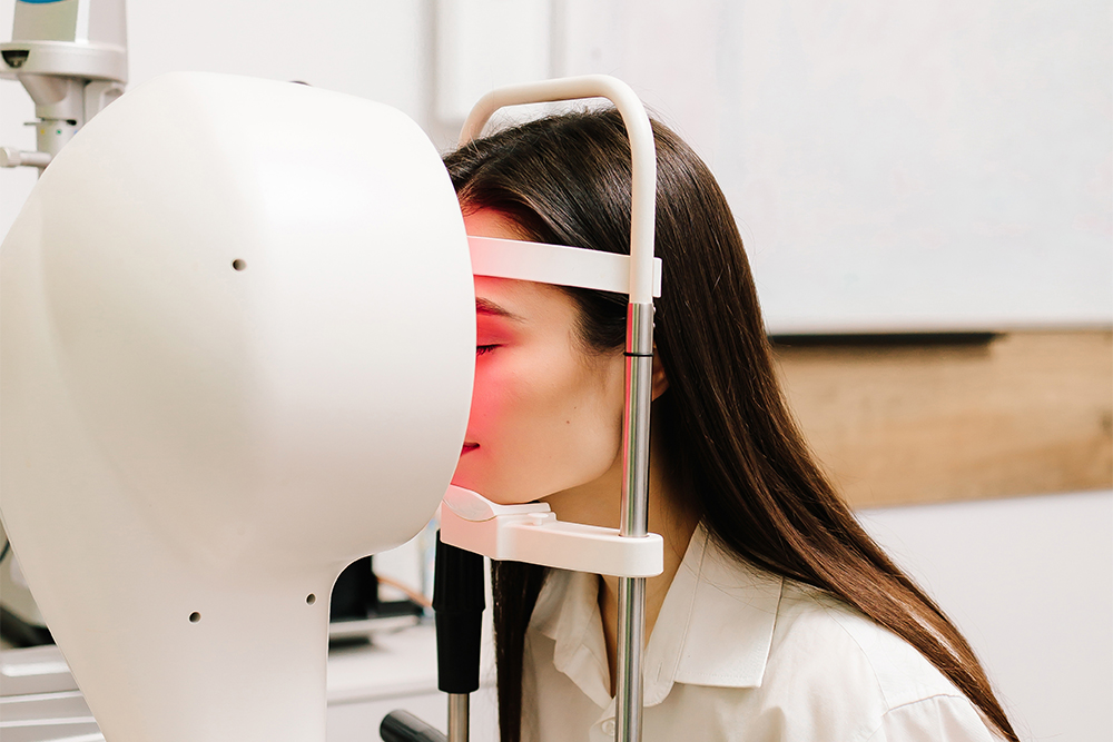
When it comes to diagnosing and managing complex vision conditions, precision matters. One of the most advanced tools is Medmont topography - a state-of-the-art technology that provides highly detailed maps of the cornea. Whether you are being evaluated for specialty contact lenses, dry eye treatment, or refractive surgery, Medmont topography plays a critical role in delivering accurate, customized care.
What Is Medmont Topography?
Medmont topography refers to corneal imaging performed with the Medmont E300 Corneal Topographer, a diagnostic device designed to capture detailed curvature measurements of the cornea.
Using advanced Placido disc technology, the system projects illuminated rings onto the cornea and analyzes their reflections. From this data, it generates a color-coded map that reveals the shape, elevation, and curvature of the cornea with exceptional accuracy.
Why Is Corneal Topography Important?
The cornea is responsible for approximately two-thirds of the eye’s focusing power. Even subtle irregularities can significantly affect vision. Medmont topography allows your eye doctor to detect and monitor:
Keratoconus and other corneal ectasias
Corneal irregularities or scarring
Post-surgical corneal changes
Astigmatism patterns
Contact lens fitting complications
Early signs of corneal distortion
Because it provides highly detailed imaging, Medmont topography often identifies changes long before they become noticeable in a routine vision exam.
How Medmont Topography Improves Contact Lens Fitting
For patients who require specialty contact lenses - such as scleral lenses, hybrid lenses, or lenses for keratoconus - precise corneal mapping is essential.
The Medmont system offers:
High-resolution curvature data
Wide corneal coverage
Repeatable, reliable measurements
Advanced software analysis
This allows your doctor to design and fit custom lenses with greater accuracy, improving comfort, stability, and visual clarity while reducing trial-and-error fittings.

In the ever-evolving world of vision correction, multifocal contact lenses have emerged as a game-changer for individuals seeking a seamless solution to address their presbyopia. As you navigate through life's various stages, your eyes may undergo changes that can impact your ability to focus clearly on objects at different distances. Multifocal contact lenses offer a convenient and effective way to combat this challenge, providing you with a versatile and comfortable option for correcting your vision.
What Are Multifocal Contact Lenses?
Multifocal contact lenses are specialized lenses designed to correct presbyopia, a condition that typically occurs as you age, making it difficult to focus on near objects. Unlike traditional single-vision lenses that correct either near or distance vision, multifocal contact lenses incorporate multiple prescriptions into a single lens. This innovative design allows you to enjoy clear vision at various distances, eliminating the need for separate reading glasses or bifocals.
How Do Multifocal Contact Lenses Work?
Multifocal contact lenses employ advanced optical technologies to provide seamless vision correction. These lenses feature distinct zones or concentric rings that correspond to different prescriptions for near, intermediate, and distance vision. As your eyes move and focus on objects at varying distances, the appropriate zone of the lens comes into play, ensuring a clear and comfortable visual experience.
There are two main types of multifocal contact lens designs:
Simultaneous Vision Design: In this design, the lens incorporates multiple prescriptions simultaneously, allowing your eyes to utilize the appropriate zone based on the viewing distance. This design can take some time to adjust to, as your brain learns to interpret the different visual inputs.
Alternating Vision Design: Also known as "segmented" or "translating" lenses, these lenses have distinct zones for near and distance vision. As you move your eyes or shift your gaze, the appropriate zone is positioned in front of your pupil, providing the necessary correction.
Eye Conditions That Multifocal Contact Lenses Can Treat
Multifocal contact lenses are primarily designed to address the following vision-related conditions:
Presbyopia: As you age, the natural lenses in your eyes lose flexibility, making it challenging to focus on nearby objects. Multifocal contact lenses can correct this age-related farsightedness, allowing you to see clearly at all distances.
Myopia (Nearsightedness): If you struggle to see distant objects clearly, multifocal contact lenses can incorporate a prescription to correct myopia, while also providing near and intermediate vision correction.
Hyperopia (Farsightedness): For individuals with farsightedness, multifocal contact lenses can offer a solution by incorporating a prescription to improve distance vision, along with near and intermediate vision correction.
Astigmatism: Some multifocal contact lenses are designed to correct astigmatism, a condition caused by an irregularly shaped cornea or lens, resulting in blurred or distorted vision.

Senior eye care is crucial for maintaining optimal vision and overall eye health as we age. As individuals grow older, they become more susceptible to age-related eye conditions. Regular comprehensive eye exams play a key role in early detection and management of these conditions, helping to preserve vision and prevent potential vision loss. By staying proactive with eye care and seeking timely professional assistance, seniors can enjoy clear, comfortable vision and ongoing eye health for years to come.
Common Eye Conditions in Seniors
As we reach our golden years, there are several eye conditions that become more prevalent. These conditions can have a significant impact on our vision and overall quality of life.
Presbyopia: This condition affects near vision, making it difficult to focus on close objects. It is a normal part of aging and is typically addressed with reading glasses or multifocal lenses.
Floaters and Flashes: As the vitreous inside the eye changes with age, it can lead to the perception of floaters (small dark spots or lines) and flashes of light. While often harmless, sudden onset of floaters and flashes may indicate a retinal tear or detachment and should be promptly evaluated.
Age-Related Macular Degeneration (AMD): AMD is a leading cause of vision loss in older adults. It affects the macula, the central part of the retina, leading to blurred or distorted central vision. There are two forms of AMD, dry AMD, and wet AMD, with the latter being more severe.
Cataracts: Cataracts are a common age-related condition characterized by the clouding of the eye's natural lens. This can lead to blurry vision, glare, and difficulty seeing in low-light conditions.
Glaucoma: Glaucoma is a group of eye conditions that damage the optic nerve, often due to elevated intraocular pressure. It can result in peripheral vision loss and, if left untreated, may lead to total blindness.
Diabetic Retinopathy: For seniors with diabetes, diabetic retinopathy is a concern. It occurs when high blood sugar levels damage the blood vessels in the retina, leading to vision problems and potential blindness if not managed.
Dry Eye Syndrome: Seniors may experience dry eye syndrome, characterized by insufficient lubrication and moisture on the surface of the eye. It can cause discomfort, redness, and fluctuating vision.

Strabismus is a relatively common condition, affecting about 2-4% of the population worldwide. It is one of the most frequent eye disorders seen in children, though it can also develop in adults due to conditions like stroke or trauma. In the United States alone, it is estimated that around 12 million people are affected by strabismus, with varying degrees of severity. Early diagnosis and treatment are essential, as untreated strabismus in children can significantly impair visual development.
What is Strabismus?
Strabismus, commonly referred to as crossed eyes, is a condition in which the eyes do not properly align with each other when looking at an object. One eye may turn in, out, up, or down, while the other eye focuses correctly. This misalignment can be constant or intermittent and may affect one or both eyes. Strabismus occurs when the muscles controlling eye movement do not work together correctly, leading to difficulty in focusing both eyes on a single point. This condition can lead to visual issues such as double vision and, in some cases, can result in amblyopia (lazy eye) if left untreated.
How Does Strabismus Develop?
Strabismus develops when there is a disruption in the coordination of the muscles that control eye movement, leading to a misalignment of the eyes. This disruption can result from various causes, including genetic factors, neuromuscular conditions, or problems with the nerves that transmit signals to the eye muscles. In some cases, it may be present at birth, while in others, it can develop later in childhood or adulthood due to conditions such as cerebral palsy, Down syndrome, or even trauma to the eye or brain.
Signs and Symptoms of Strabismus
The primary symptom of strabismus is the visible misalignment of the eyes. However, there are other signs and symptoms that may indicate the presence of this condition:
Blurred or double vision
Headaches or eye strain
Difficulty with depth perception and spatial awareness
Reduced visual acuity in one eye
Frequent eye rubbing or squinting
Tilting or turning the head to compensate for the misalignment

All Eye
Care Services

Keep
In Touch

Kissel Eye Care
Neurovision Rehab Center of PA
© 2026 Kissel Eye Care. All Rights Reserved. Accessibility Statement - Privacy Policy - Sitemap


